Your Body Knows Before You Do: The Science of Emotions Written in Signals
In our previous article, we explored how emotions arise from the perspective of psychological mechanisms and brain structures. We discovered that emotions are not vague and abstract mental feelings, but rather concrete physiological responses that manifest through bodily signals in very real ways.
Whenever we feel nervous, excited, afraid, or angry, the brain immediately sends commands to the body. These instructions are rapidly transmitted via the nervous system and hormonal pathways, regulating our heart rate, breathing, sweating, muscle tension, and other physiological states. In fact, our bodies often react before we are even consciously aware of our emotions.
In today’s article, we’ll continue shifting our lens from the brain to the entire body, to see how exactly emotions are written into our physiological states.
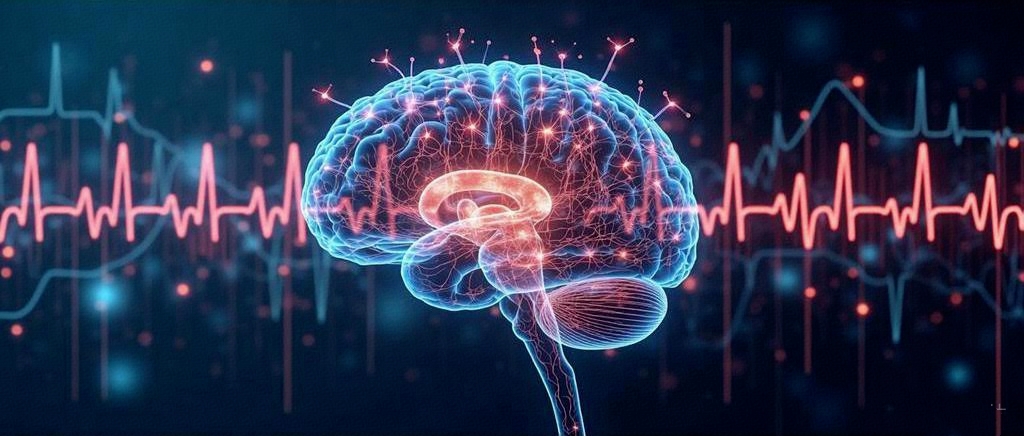
Among the many physiological signals, two have been repeatedly studied in neuroscience and are most indicative of emotional fluctuations: ECG (electrocardiogram) and EEG (electroencephalogram). These represent the two key centers of emotional regulation in the body—the heart and the brain.
Emotional Clues Hidden in Heartbeats: ECG
The heart is not only a life-sustaining pump but also a core participant in emotional processes. It is closely connected to the brain through a complex, high-speed bidirectional neural highway known as the Heart-Brain Axis. This axis, consisting of the vagus nerve and other autonomic pathways, ensures that every emotional fluctuation in the brain leaves a trace in the heart’s rhythm.
When we experience emotional stress—fear, anger, tension—the limbic system in the brain, especially the amygdala and prefrontal cortex, sends signals to the autonomic nervous system. These signals modulate heart rhythms, vascular dilation, and other physical responses. In return, the state of the heart sends feedback to the brain, influencing emotional appraisal and behavioral decisions.
Thus, emotions are not merely experiences in the brain—they are also deeply inscribed into our heart’s beating patterns.
ECG: Interpreting Emotions Through Cardiac Signals
The ECG is the primary tool for recording heart activity. Every time the heart beats, cardiac muscle cells generate tiny electrical fluctuations. ECG uses electrodes to detect these signals and create a dynamic “activity map” of the heart.
One key metric in emotion research is Heart Rate Variability (HRV)—the variation in the time interval between two consecutive heartbeats (the R-R interval).
HRV: A Physiological Barometer of Emotional Regulation
Interestingly, HRV is not about having a steady heartbeat. On the contrary, higher variability often indicates a healthier body and more flexible emotional regulation.
- High HRV suggests the sympathetic and parasympathetic systems can rapidly shift, indicating that the brain is effectively managing emotional stress. This is commonly observed after relaxation, meditation, or mindfulness practice.
- Low HRV is often found in individuals with chronic stress, anxiety disorders, depression, or fatigue, reflecting weakened self-regulation.
From a neuroscience standpoint, HRV offers a feedback window into the coordination between the prefrontal cortex (which governs self-control) and the limbic system (which handles emotions). It helps us understand whether our emotional brain is overactive or whether our cortical brain is still in command.
Due to its strong association with emotional health, HRV is widely applied in areas such as:
- Mental health assessment: Continuous HRV monitoring can help detect early declines in emotional regulation, aiding diagnoses of anxiety, depression, and more.
- Stress monitoring: Many wearable devices (like Apple Watch or Fitbit) now include HRV metrics to evaluate users’ stress levels.
- Affective AI: HRV has become a core physiological feature in emotion-aware systems, powering applications such as personalized music recommendation or therapeutic interventions.
Emotional Waves in the Brain: EEG
If ECG is an echo of our emotional state in the body, then EEG is a window directly into the brain—the very center of emotion.
Our brain is composed of tens of billions of neurons that communicate via electrical signals. When groups of neurons activate simultaneously, they create subtle electrical fields detectable on the scalp. EEG captures these group discharges using electrodes placed on the scalp and visualizes brain activity in real time.
Though these signals are minuscule—usually in microvolts (μV)—they form the physical foundation of our cognition, emotion, and consciousness.
Different Brain Rhythms Encode Different Emotions
EEG signals are categorized into frequency bands, each associated with specific psychological and emotional states:
- Delta waves (0.5–4 Hz): Dominant during deep sleep but may be abnormally elevated in depressive states.
- Theta waves (4–8 Hz): Linked to relaxation, meditation, emotional imagery, and dream-like states; also connected to anxiety.
- Alpha waves (8–13 Hz): Prominent during relaxed wakefulness, especially when eyes are closed—often considered a sign of calm.
- Beta waves (13–30 Hz): Associated with active thinking, alertness, anxiety, and tension.
- Gamma waves (30+ Hz): Possibly linked to higher cognitive processes, emotional perception, and empathy (though still under research).
These brainwaves rarely occur in isolation. They activate in patterns depending on our mental state. For instance, under stress, alpha waves decrease while beta waves increase; during meditation, the pattern reverses.
Frontal Asymmetry: The Neural Signature of Emotional Tendencies
Beyond frequency bands, EEG also reveals differences in activation across brain regions—especially the frontal lobe.
A well-studied phenomenon is frontal asymmetry:
- Greater left frontal activity is associated with positive emotions, proactive motivation, and approach behavior.
- Greater right frontal activity often corresponds to anxiety, depression, or avoidance behavior.
This asymmetry, known as the Frontal Asymmetry Theory, is already being applied in diagnostic tools and emotional training interventions.
EEG’s non-invasive nature and high temporal resolution (on the order of milliseconds) make it valuable in areas such as:
- Emotional disorder assessment: Aiding diagnoses of anxiety, depression, or autism spectrum conditions through EEG markers.
- Meditation and emotional training: EEG is integrated into feedback systems for mindfulness and concentration.
- Emotion-aware AI: Combining EEG and machine learning to identify user emotions, enabling personalized therapy or immersive experiences.
- Brain-Computer Interfaces (BCI): EEG is being explored for interpreting emotional intent and enabling emotion-based communication—particularly in autism therapy.
EEG brings emotional activity from the brain into view, helping bridge the gap between our inner world and external behaviors.
What Other Body Signals Reveal Emotions?
Beyond ECG and EEG, the body offers many other valuable indicators of emotion. Though often overlooked, these signals reflect how our body quietly responds to both environment and inner states.
Galvanic Skin Response (GSR)
When we are anxious, scared, or excited, sweat glands become more active—even without visible perspiration—making the skin more conductive. GSR measures this change in conductivity to infer arousal levels.
It is closely linked to sympathetic nervous system activity and serves as a key indicator of stress and alertness.
Breathing Patterns and Frequency
Emotions influence breathing directly:
- Anxiety and fear: fast, shallow breathing
- Anger: irregular, chest-centered breathing
- Calmness: slow, deep breaths
Respiratory belts or breath sensors can track these changes in real time, offering insight into emotional states and supporting anxiety intervention training.
Electromyography (EMG)
Emotions—especially anger or tension—cause subtle muscle contractions, such as frowning, clenching teeth, or pursing lips. EMG captures these muscle signals.
This is particularly useful in situations where facial expressions are hard to detect (e.g., video calls), offering a more precise emotional reading.
Body Temperature, Blood Pressure, and Pupil Size
- Emotional arousal can raise local body temperature (e.g., cheeks or earlobes).
- Blood pressure reflects long-term emotional stress.
- Pupil diameter varies with arousal and can indicate emotional intensity.
Though less prominent than ECG and EEG, these signals complement each other like puzzle pieces, forming a rich emotional portrait.
We’re Finally Learning to Listen to Our Bodies
Have you ever had a moment like this:
- Your heart suddenly races, for no apparent reason.
- You sit quietly, yet your palms start to sweat.
- A song plays, and tears well up without warning.
This isn’t emotional weakness. It’s your body speaking.
Emotions have never been intangible illusions—they are physiological states that can be recorded, measured, and understood. ECG and EEG allow us to scientifically decode this language of the body. And the other signals—GSR, respiration, EMG, temperature—are emotional signatures written across your being.